
05
Dec
Nervous tissue is composed of two types of cells, neurons and glial cells. Astrocytes, oligodendrocytes, and microglial cells (figure 1.4a—c).
Color the neuron and neuroglial cells. For over acentury, it was believed that they did not play any role in neurotransmission. What are 6 types of glial cells? Color the neuron and neuroglia.
Color the neuron and neuroglial cells oligodendrocytes (purple) astrocyte (green) body of neuron (blue) ependymal cells (orange) myelin sheaths. They are responsible for the computation and communication that the nervous system provides. There are no instructions, students must identify each of the types of glial cells:
Name lina color the neuron and neuroglial cells due by 3 / s fluid filled cavity of brain capillary oligodendrocytes Anatomy & physiology oligodendrocytes (purple) astrocyte (green) ependymal cells (orange) body of neuron (blue) myelin sheaths (pink) capillary (red) microglial cells (yellow) nodes or ranvier and the axon (brown) Color the neuron and neuroglia 0914 neuroglial cells astrocyte medical images for powerpoint | powerpoint slide.
Oligodendrocytes (purple) astrocyte (green) ependymal cells (orange) body of neuron (blue) myelin sheaths (pink) capillary (red) microglial cells (yellow) nodes of ranvier and the axon (brown) questions 1) describe the function of the following parts: Main functions of glial cells: Image of nervous tissue showing cells of the neuroglia, including the neuron, astrocytes, oligodendrocytes, and microglia.
Oligodendrocytes (purple) astrocyte (green) ependymal cells (orange) body of neuron (blue) for each of the cells above, color the nucleus a darker shade of purple, green, blue, orange. They compose a rich support system that is essential to the operation of nervous tissue and the nervous system. Astrocytes, oligodendrocytes, and microglial cells (figure 1.4a—c).
This coloring worksheet is intended for anatomy students who have already been introduced to neurons and neuroglial cells. Neurons are the primary type of cell that most anyone associates with the nervous system. Unlike neurons, glial cells do not have axons, dendrites, or conduct nerve impulses.
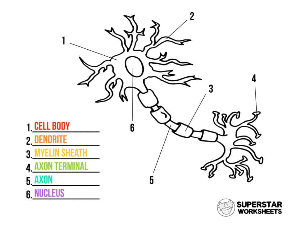
Use this video to help visualize the neuroglial cells in the color the neuron and neuroglial cells worksheet. Labeled neuron (labeled neuron.jpg) use this to color and label the neuron. 12/3/2020 1 color the neuron and neuroglial cells color the following:
Neuroglia are typically smaller than neurons and are about three times more. Nervous tissue is composed of two types of cells, neurons and glial cells. There are six types of neuroglia, each with different functions:
What cell is found in the cns and connects neurons to blood vessels and form scar tissue? That idea is now discredited; Body of neuron (blue) ependymal cells (orange) for each of the cells above, color the nucleus a darker shade of purple, green, blue, orange myelin sheaths (pink) capillary (red) microglial cells (yellow) nodes or ranvier and the axon (brown)
Glial cells are far more numerous than neurons and, unlike neurons, are capable of mitosis. Oligodendrocytes, astrocyte s, microglial cells, , and ependymal cells. I even have a neuron plushie to go along with this lesson and use toilet paper rolls to model the myelin sheath.
Glial (neuroglial) cells do not conduct nerve impulses, but, instead, support, nourish, and protect the neurons. They are electrically active and release chemical signals to target cells. Color according to directions and answer basic questions about the anatomy of a neuron.
There are three types of glial cells in the mature central nervous system: Astrocytes, which are restricted to the brain and spinal cord, have elaborate local processes that give these cells a starlike appearance (hence the prefix “astro”). View nerve cells coloring.pdf from bio ap at frankel jewish academy of metro.
Color the neuron and neuroglial cells name: To surround neurons and hold them in place, to supply nutrients and oxygen to neurons, to insulate one neuron from another, and to destroy pathogensand remove dead neurons. Name_ color the neuron and neuroglial cells oligodendrocytes.
In the past, students would label a nerve cell and color neuroglia cells using paper handouts to learn the structures of the neuron and the neuroglia (supporting cells). Myelin sheaths (pink) capillary (red) microglial cells (yellow) nodes or ranvier and the axon (brown) what is the function of:
Previous post
Columbus ohio swing clubsNext post
Color remover vs bleach